Pulmonary Function Tests (PFTs)
Duration: 29:55
Published On Apr 14, 2017
This video presents the lung function tests.
In this video we will learn about :
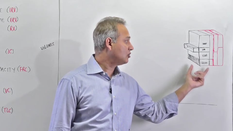
1. Normal lung volume capacities.
2. Lung flow volume graph.
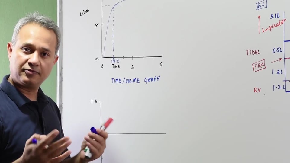
3. Time volume graph.
3. Changes in restrictive and obstructive lung diseases.
4. Extra pulmonary air way compression.
5. Blunting of the graph.
Following answers are created by ChatGPT. Occasionally the answer may be harmful, incorrect, false, misleading, incomplete, or limited in knowledge of world. Please contact your doctor for all healthcare decisions. Also, double check the answer provided by the AI below.
Faculty
In addition to the presenter, following authors may have helped with the content writing, review, or approval:
CME, CE, CEU and Other Credit Types:
ACCME Accreditation Statement
The DrBeen Corp is accredited by the Accreditation Council for Continuing Medical Education (ACCME) to
provide continuing medical education for physicians.
AMA Credit Designation Statement
The DrBeen Corp designates this enduring material for a maximum of 0.5 AMA PRA Category 1
Credits™.
Physicians should claim only the credit commensurate with the extent of their participation in the
activity.
Disclosure Information
In accordance with the disclosure policies of DrBeen Corp and the ACCME (Accreditation Council for
Continuing Medical Education), we are committed to upholding principles of balance, independence,
objectivity, and scientific rigor in all of our Continuing Medical Education (CME) and Continuing
Education (CE) activities. These policies include the careful management and mitigation of any relevant
financial relationships with organizations that are not eligible.
All members of the Activity Planning Committee and presenters have disclosed their relevant financial
relationships. The DrBeen Corp CE Committee has thoroughly reviewed these disclosures and determined
that these relationships are not deemed inappropriate in the context of their respective presentations.
Additionally, they are found to be consistent with the educational objectives and the integrity of the
activity.
| Faculty | Disclosures |
|---|---|
| Author declares no conflict of interest. |
Instructors
Respiratory Physiology
 0.75 CME
Dead Spaces and Ventilation Rates
40:43
Dr. Mobeen Syed
 0.75 CME
Introduction to Gas Exchange
44:17
Dr. Mobeen Syed
0.75 CME
Dead Spaces and Ventilation Rates
40:43
Dr. Mobeen Syed
 0.25 CME
Lung Capacities and Volumes
21:57
Dr. Mobeen Syed
 0.12 CME
Abnormal Breathing Patterns
07:13
Dr. Mobeen Syed
 0.22 CME
Quiet Breathing Cycle
13:04
Dr. Mobeen Syed
 0.50 CME
Muscles of Breathing
33:43
Dr. Mobeen Syed
 0.75 CME
Forces on the Lung System
39:57
Dr. Mobeen Syed
 0.25 CME
Forces on the Lung System Review
16:28
Dr. Mobeen Syed
 0.22 CME
Pneumothorax
12:51
Dr. Mobeen Syed
 0.25 CME
Positive Pressure Ventilation
16:45
Dr. Mobeen Syed
 0.50 CME
Lung Compliance
32:47
Dr. Mobeen Syed
 0.50 CME
Pulmonary Pressure and Flow Resistance
26:32
Dr. Mobeen Syed
 0.25 CME
Fick's Principle
20:49
Dr. Mobeen Syed
 0.50 CME
Transport of Gases
25:05
Dr. Mobeen Syed
 0.75 CME
Oxygen Dissociation Curve
41:23
Dr. Mobeen Syed
 0.25 CME
CO2 Transport
15:05
Dr. Mobeen Syed
 0.14 CME
Bohr Effect
08:25
Dr. Mobeen Syed
 0.50 CME
Ventilation Perfusion Ratio (V/Q)
29:43
Dr. Mobeen Syed
 0.14 CME
Haldane Effect
07:58
Dr. Mobeen Syed
0.50 CME
Pulmonary Function Tests (PFTs)
29:55
Dr. Mobeen Syed
 0.50 CME
Regulation of Breathing
32:41
Dr. Mobeen Syed
 0.25 CME
Protective Mechanisms against Pneumonia
19:44
Dr. Mobeen Syed
Write A New Comment
2 Comments
gkikat@*.com
Feb 23 2019, 7:43 pm
Excuse me, can I add one more question?
In a patient with obstructive disease, let's say asthmatic patient, the airway diameter is smaller, so how is he going to breath in the same amout of air. Is it a matter of time?
I mean, he would breath the same amount of air as a normal person, but in more time? (with decreased slope factore in a volume-time diagram for instance ? )
gkikat@*.com
Feb 23 2019, 7:16 pm
Dear Dr. Mobeen,
I would like to ask 2 questions.
1) In a patient with emphysema, there are dilated alvioli and "trapped air". So there is increased residual volume. So far so good, BUT, since the alvioli membranes are destroyed and on top of that there is trapped air, shouldn't that mean that the alvioli of the damadged area are less compliant and thus they can take in less air. ? So why is there a normal inspiration volume? I understand the normal FVC only because of an increased RV. But is the IV normal? Can the patient with emphysema inspire the same amount of air as with a normal person?
2) In the restrictive diseases, you mentioned that the scar is pulling airways open, (I understand it as pulling the airways out and they open ). Compliance is less. So IV is less. But since the scar is pulling outwards, why do they tend to collapse?, why is the recoil force intact ? (since there is a force pulling in the opposite direction of that of collapsing ) and by extention why is the expiration process normal?
Thank you very much doctor. You are really making us better professionals. Can't thank you enough for all of your lectures (And this is a genuine expression of appreciation)