Fundamentals of Chest X-Ray Interpretation
Duration: 1:53:47
Published On Jun 2, 2017
Chest X-Ray is a routine clinical investigation. It is important for healthcare professionals to be comfortable approaching and interpreting it. In this webinar, Dr. Alikhan shares his excellent method to identify key elements that are frequently encountered on the chest x-ray.
In this video the learner will get to know about:
- Normal CXR appearance and identification of anatomical landmarks.
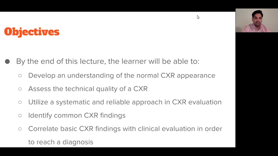
- Assessment of the technical quality of CXR.
- Systematic approach to CXR evaluation and interpretation.
- Understanding of deviations in structures and associated pathologies.
- Identification of common CXR findings or pathologies.
- Clinical correlation of CXR findings and establishment of provisional diagnosis.
- Discussion of some common clinical scenarios on radiological findings.
Presented by Dr. Mir Alikhan
Following answers are created by ChatGPT. Occasionally the answer may be harmful, incorrect, false, misleading, incomplete, or limited in knowledge of world. Please contact your doctor for all healthcare decisions. Also, double check the answer provided by the AI below.
Faculty
In addition to the presenter, following authors may have helped with the content writing, review, or approval:
CME, CE, CEU and Other Credit Types:
ACCME Accreditation Statement
The DrBeen Corp is accredited by the Accreditation Council for Continuing Medical Education (ACCME) to
provide continuing medical education for physicians.
AMA Credit Designation Statement
The DrBeen Corp designates this enduring material for a maximum of 2 AMA PRA Category 1
Credits™.
Physicians should claim only the credit commensurate with the extent of their participation in the
activity.
Disclosure Information
In accordance with the disclosure policies of DrBeen Corp and the ACCME (Accreditation Council for
Continuing Medical Education), we are committed to upholding principles of balance, independence,
objectivity, and scientific rigor in all of our Continuing Medical Education (CME) and Continuing
Education (CE) activities. These policies include the careful management and mitigation of any relevant
financial relationships with organizations that are not eligible.
All members of the Activity Planning Committee and presenters have disclosed their relevant financial
relationships. The DrBeen Corp CE Committee has thoroughly reviewed these disclosures and determined
that these relationships are not deemed inappropriate in the context of their respective presentations.
Additionally, they are found to be consistent with the educational objectives and the integrity of the
activity.
| Faculty | Disclosures |
|---|---|
| Author declares no conflict of interest. |
Instructors
Respiratory Medicine
 0.75 CME
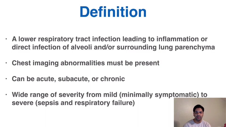
Lower Respiratory Tract Complications
45:21
Mir Alikhan, MD
 0.25 CME
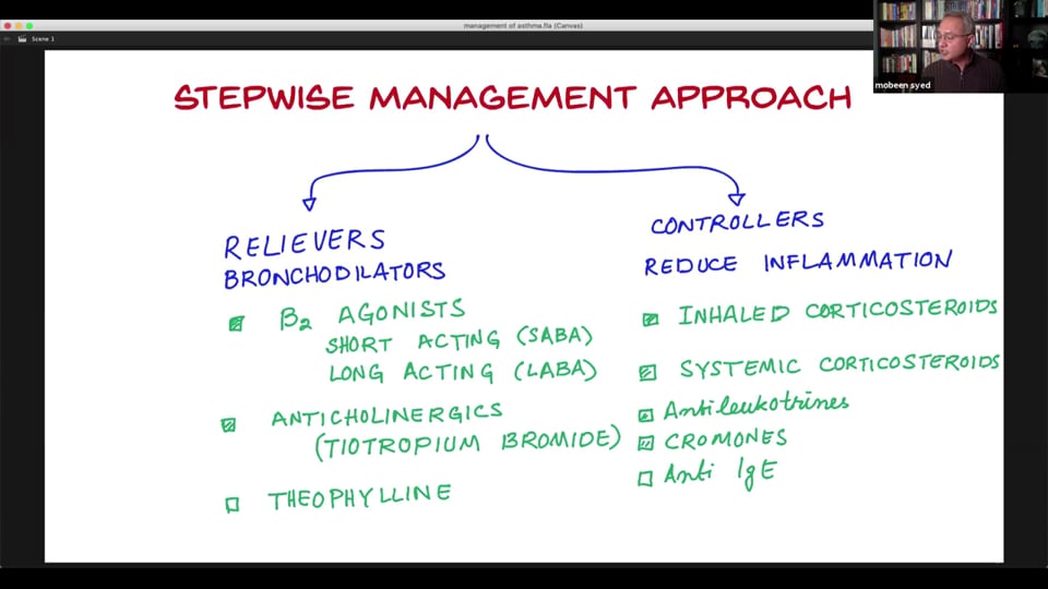
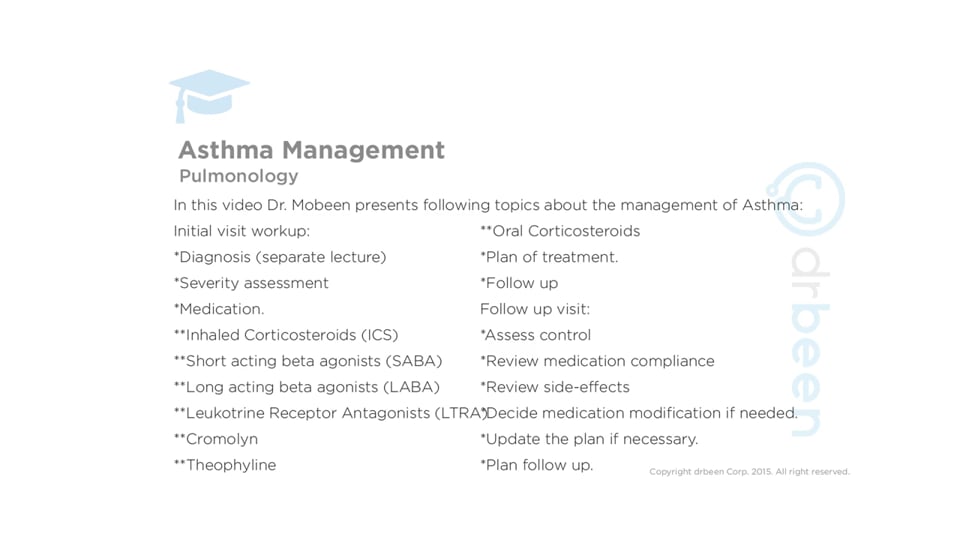
Management of Asthma Part 3
18:00
Dr. Mobeen Syed
 0.75 CME
Management Of Asthma Part 1
44:08
Dr. Mobeen Syed
 0.25 CME
Clinical Applications of Anti-Histamine Drugs (Part 2)/Common Cold
19:41
Ahmed Zaafran, MD
 0.75 CME
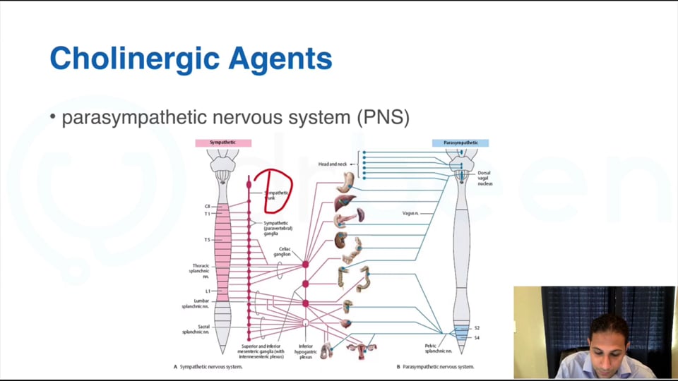
Clinical Application of Cholinergic Drugs/Cholinergic System
51:32
Ahmed Zaafran, MD
 0.75 CME
Asthma Types and Clinical Pathophysiology
52:09
Dr. Mobeen Syed
 0.75 CME
A Clinical Approach to Asthma
38:26
Ahmed Zaafran, MD
 0.25 CME
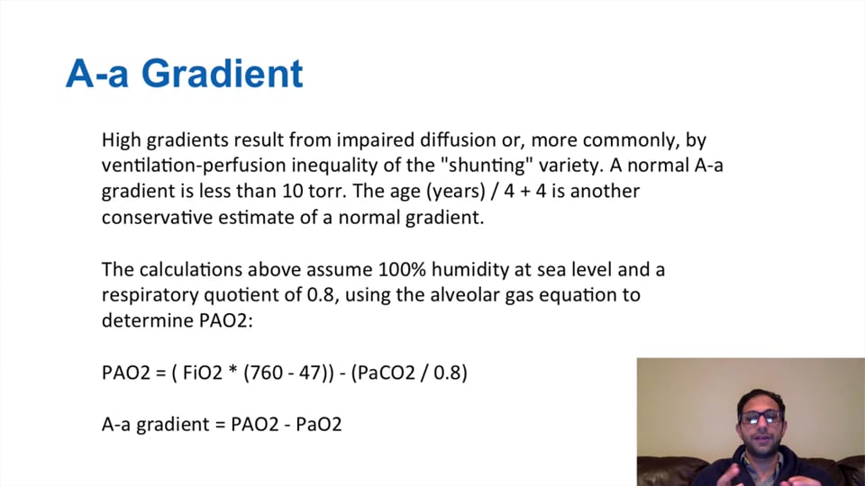
A Clinical Approach to Hypoxemia/Blood Gases
17:54
Ahmed Zaafran, MD
 0.25 CME
Clinical Applications of Anti-Histamine Medications (Part 1)/Common Cold
19:06
Ahmed Zaafran, MD
 0.75 CME
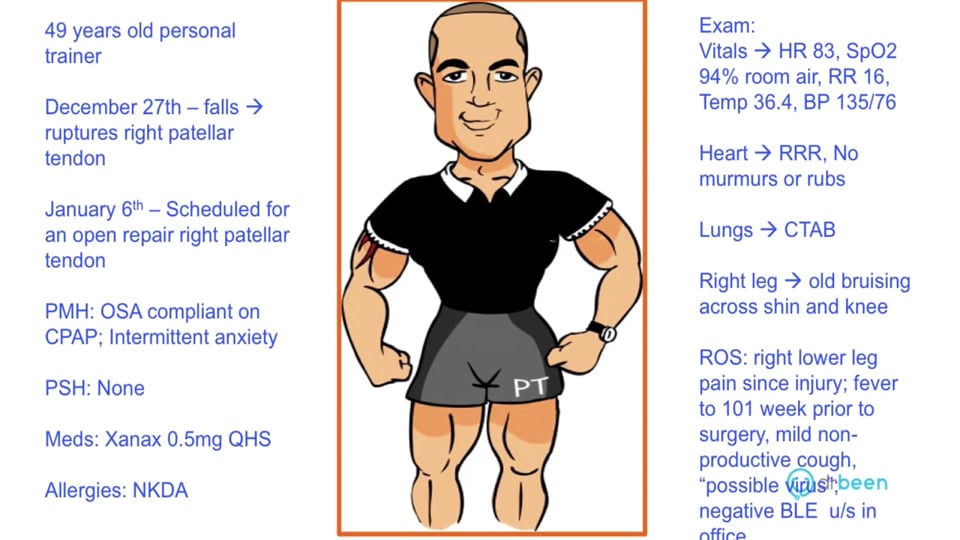
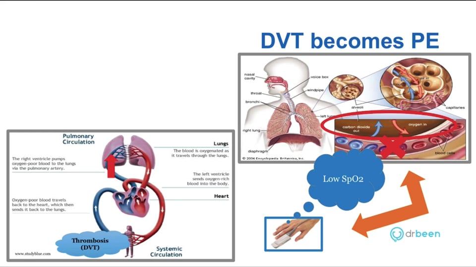
Pulmonary Embolism - Case Discussion and Treatment Approach
40:38
Ana Crawford M.D., M.Sc.
 0.25 CME
Pulmonary Embolism - Definition, Pathophysiology, and Presentation
17:40
Ana Crawford M.D., M.Sc.
 0.50 CME
Clinical Application of Respiratory Drugs (Part2) / Asthma
22:41
Ahmed Zaafran, MD
 0.25 CME
Common Clinical Cases, Acid-Base Disorders
21:11
Ana Crawford M.D., M.Sc.
 0.50 CME
Acid Base Disorders
32:02
Ana Crawford M.D., M.Sc.
 0.75 CME
ARDS - Management of Patients
38:03
Ana Crawford M.D., M.Sc.
 0.25 CME
ARDS - Pathophysiology and Clinical Presentation
17:52
Ana Crawford M.D., M.Sc.
 0.50 CME
Clinical Applications of Respiratory Drugs (Part 1)/ Asthma
26:05
Ahmed Zaafran, MD
 0.50 CME
Asthma Management
33:30
Dr. Mobeen Syed
 0.50 CME
Asthma Classification
29:58
Dr. Mobeen Syed
 0.50 CME
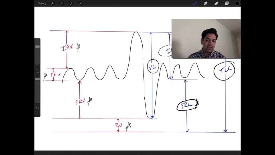
Interpretation of Pulmonary Function Tests
23:28
Mir Alikhan, MD
2.00 CME
Fundamentals of Chest X-Ray Interpretation
1:53:47
Mir Alikhan, MD
0.75 CME
Lower Respiratory Tract Complications
45:21
Mir Alikhan, MD
 1.00 CME
Pulmonary Vascular Disease
57:06
Mir Alikhan, MD
 0.25 CME
Upper Respiratory Tract Infections
17:46
Mir Alikhan, MD
Write A New Comment
2 Comments
momofben@*.com
Apr 25 2026, 5:34 am
Video is extremely difficult to hear
citypkctmc@*.com
May 23 2025, 4:51 am
Indirect signs on a chest X-ray that might be seen in PE include: Hampton's hump: A peripheral, wedge-shaped opacity in the lung, often seen at the lung periphery and may indicate pulmonary infarction. Westermark's sign: Regional oligemia (reduced blood flow) in the lungs, characterized by areas of increased lucency. Fleischner sign: Enlargement of the central pulmonary artery, often seen with a large clot in the pulmonary artery. Knuckle sign: A sudden tapering of the pulmonary artery, suggesting a clot in the artery. Palla sign: Enlargement of the right descending pulmonary artery. Atelectasis: Collapse of lung tissue, often seen in areas of infarction. Pleural effusion: Fluid accumulation in the space between the lungs and chest wall. Prominent central pulmonary artery: Fleischner's sign is an enlarged central pulmonary artery.