This video presents the histophysiology of the artries capillaries and veins.
STUDY NOTES:
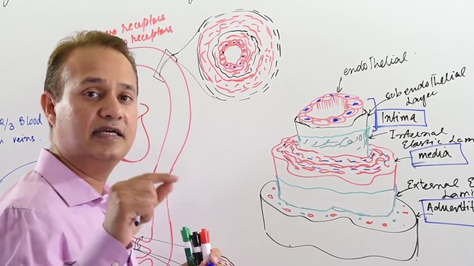
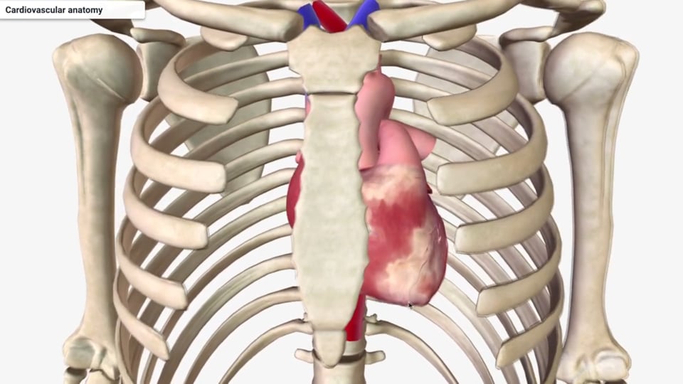
HISTOLOGY OF BLOOD VESSELS
This lecture discusses about the structure, function and the differences in between the different components of the vasculature (arteries, arterioles, capillaries, venules, veins). Before delving into the details about individual components of the vasculature, an understanding about the general structure of the blood vessel will be discussed. A standard blood vessel is composed of three layers, which are discussed as following:
In various types of vessels these above mentioned layers would be either present or absent. If present, the composition and thickness of these layers can slightly vary from one vessel type to another.
TYPES OF VASCULATURE
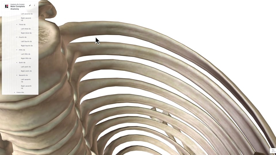
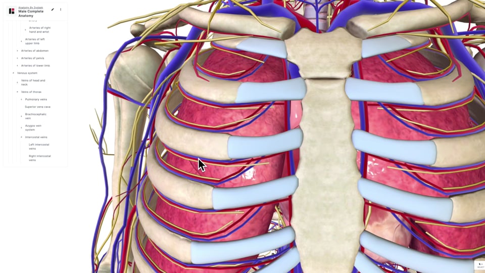
ARTERIES conduct blood away from the heart towards the tissues. Arteries normally carry oxygenated blood away from the heart. However, exceptions to this rule are the pulmonary arteries and the umbilical arteries which carry deoxygenated blood away from the heart.
Elastic arteries are larger in size and have a relatively thick tunica media. Examples of elastic arteries include the pulmonary arteries and aorta plus its 3 major branches (left brachiocephalic, right common carotid & the right subclavian arteries). Tunica media is so large in the case of aorta, that it has 50 layers of alternating elastic and smooth muscle fibers present in an alternating circular pattern. Elastic arteries are also referred to as the conducting arteries. Their main function is to receive high pressure blood from the heart and distend outwards keeping the blood there before they slowly return to their normal caliber and gently push the blood forward as the heart enters diastole phase. This is called the windkessel effect and it prevents any turbulence of the high pressure blood which leaves the heart. Hence, the elastic component allows the elastic arteries to recoil appropriately and serve their function as conducting vessels.
Endothelium is the innermost layer of the tunica intima and this is surrounded by the subendothelial layer. In case of arteries, the subendothelial layer has a lot of elastic and smooth muscle fibers. Subendothelial layer is the part of the tunica intima that is most commonly involved in atherosclerosis. Atherosclerosis is an intimal disease which results in formation of atheromatous plaques within the subendothelial layer of the tunica intima. Atherosclerosis most commonly affects the medium and large sized arteries. This plaque formation tends to decrease the luminal size of the vessels and consequently the blood which flows across them. Arteries have abundant smooth muscle and elastic fibers in their tunica media, which gives them the recoil capability. Tunica adventitia forms the outermost layer. Small vessels called the vasa vasorum are located within the tunica adventitia, and they function to vascularize the wall of the walls of the elastic arteries.
Baroreceptors and chemoreceptors are present within the walls of carotid sinus and the arch of aorta. Baroreceptors are formed by the thinning of the wall of these arteries. This allows the increased blood pressure to have a more pronounced effect in distending the arterial wall in this region where the baroreceptors are located. There are nerves located in the adventitia of the baroreceptor region which sense this stretch of the vessel and send impulses if there’s a significant rise or drop in blood pressure. Similarly there are chemical receptors that are also present within the muscular wall of the arch of aorta. These chemoreceptors are formed by Glomus type 1 cells which are situated within the walls of the arch of aorta. These Glomus type 1 cells have channels for oxygen, carbon dioxide and hydrogen ions; this allows them to act as sensory receptors for the monitoring the concentrations of oxygen & carbon dioxide plus the pH of the arterial blood. Glomus type 1 cells are supported by the satellite receptors. These receptors respond to hypoxia, hypercapnia (increased arterial CO2 levels) and the pH changes in the arterial blood. Any derangements of these above mentioned parameters result in firing of the glomus cells thereby activating the corresponding control mechanism to ensure homeostasiswithin the body.
Muscular arteries arise from the larger elastic arteries. They are also referred to as the conducting arteries and they supply blood to various organs of the body. These muscular arteries mainly respond to sympathetic/adrenergic control because of the presence of alpha 1and beta 2 receptors. Sympathetic stimulation is responsible for the vasoconstriction effect in these arteries. They do have an endothelial and subendothelial layer. The smooth and elastic fiber layer in the subendothelium is not that dense. Their tunica media is composed of 3 to 10 layers of smooth muscle and elastic fibers. Outermost layer is formed by the tunica adventitia. Muscular arteries are also referred to as resistance vessels. This is because they exhibit vasoconstriction upon sympathetic stimulation; hence they can change their diameter. Upon decreasing their diameter, they can decrease the forward flow hence they are called resistance vessels. The volume of blood in the muscular arteries is referred to as stressed volume, since these arteries are always compressing upon the blood inside them to help maintain the blood pressure.
The ARTERIOLES form the terminal arterial vessels and form a connection between the arteries and the capillaries. Arterioles form the major resistance site of the systemic vasculature and therefore, they are responsible for maintaining the blood pressure. The arterioles are also referred to as the functional sphincters of the arterial system. Within the tunica intima, they do have an endothelial and subendothelial layer. Subendothelial layer is not that dense in elastic and muscle fibers. Their tunica media has 1 to 3 layers of smooth muscle and elastic fibers. This tunica media responds to sympathetic and parasympathetic stimulation. Sympathetic stimulation achieves vasoconstriction of the arterioles and results in a decreased forward flow through them. In contrast, the parasympathetic stimulation achieves vasodilatation of the arterioles, which results in an increased forward flow across the arteriole.
In the case of skin, gastrointestinal system and the renal system; the sympathetic stimulation of their arterioles is modulated via the alpha-1 receptors. The arterioles leading to the skeletal muscles are controlled by beta 2 receptors.
CAPILLARIES have the smallest caliber in terms of luminal diameter. Though the capillaries have a small diameter, they have the greatest cross-sectional area amongst all the vasculature. The total cross-sectional area of all the capillaries in the body can add up 2/3 of a meter. Also they have the largest surface area relative to all the blood vessels in the body. The tunica intima of capillaries is only composed of an endothelium and a basement membrane. The endothelium is formed by a single layer of endothelial cells which allows them to function as sites of exchange. Tunica media and adventitia are absent in the capillaries. Capillaries function as the exchange site of the vasculature because they exhibit selective permeability of substances across their walls. This permeability allows diffusion of oxygen, nutrients, signaling molecules and water from the blood to the tissues. Carbon dioxide and waste products diffuse in the opposite direction from the tissues into the capillaries.
When the capillaries merge together they end up forming VENULES. The venules have an endothelium and a broken subendothelium but they lack tunica media and adventitia. They are normally collapsed and have a low blood pressure. Venules are also involved in exchange of metabolites with the surrounding tissue. Another important function of venules is diapedesis which involves cellular trafficking across the wall of the venules. This is made possible by endothelial contraction which allows gaps to appear within the endothelium of the postcapillary venules. This diapedesis allows venules to function as sites of leukocyte extraction into the tissues. Inflammatory mediators released from the inflamed tissues allow leukocyte recruitment and via diapedesis leukocyte trafficking occurs into the tissue space. This allows leukocytes to take active part at the site of inflammation.
VEINS are formed when the venules merge together. The veins function as a reservoir of the blood since 2/3rd of the total blood volume is present in the veins at any given time. Veins transport blood away from the tissues and return it to the heart. Veins normally carry deoxygenated blood, except the pulmonary veins in adults and the umbilical vein during fetal life which bring back oxygenated blood to the heart from the lungs and the placenta respectively. Veins as compared to arteries are thin walled, more compliant & distensible, have a greater luminal diameter and have valves which prevent the backward flow of blood. Veins lack the elastic and muscular component hence when the blood backflows under the influence of gravity, the valves tend to approximate and prevent the retrograde flow of blood. In contrast to venules, larger veins have more defined endothelium & subendothelium and they have a tunica media as well.
In this lecture Dr. Mobeen will discuss:
1. Histology of blood vessels. (0:44)
2. Structure of elastic arteries. (2:13)
3. Structure of simple arteries and their properties. (7:20)
4. Arterioles: Structure and function. (9:00)
5. Capillaries: (10:38)
6. Venules. (12:15)
7. Structure of veins and their properties. (12:50)
Presented by Dr. Mobeen Syed
No credit card information needed.


Write A New Comment
0 Comments