Gram staining
Duration: 10:05
Published On Apr 14, 2017
Mechanism of Gram staining - Dr. Mobeen Syed
Put bacteria on plate (if from another plate then dilute agar first);
-
Warm the plate to fix bacteria;
-
Add gram stain; 20-30 seconds; wash it;
-
Add iodine (mordant)
-
Add drops of alcohol, to wash out the gram negative bacteria’s color - 20 - 40 seconds;
-
Put safranin (counter stain) 20-40 seconds
-
What Happens?
- * Gram positive (+ive) bacteria turns purple
- * Gram negative(-ive) bacteria turns red
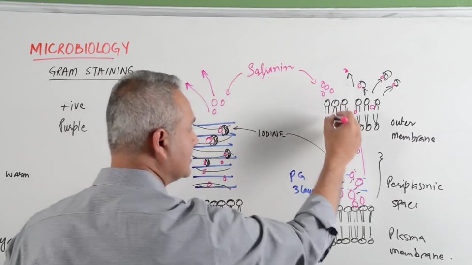
Go to TC 2:57 in the video for illustration of how it happens:
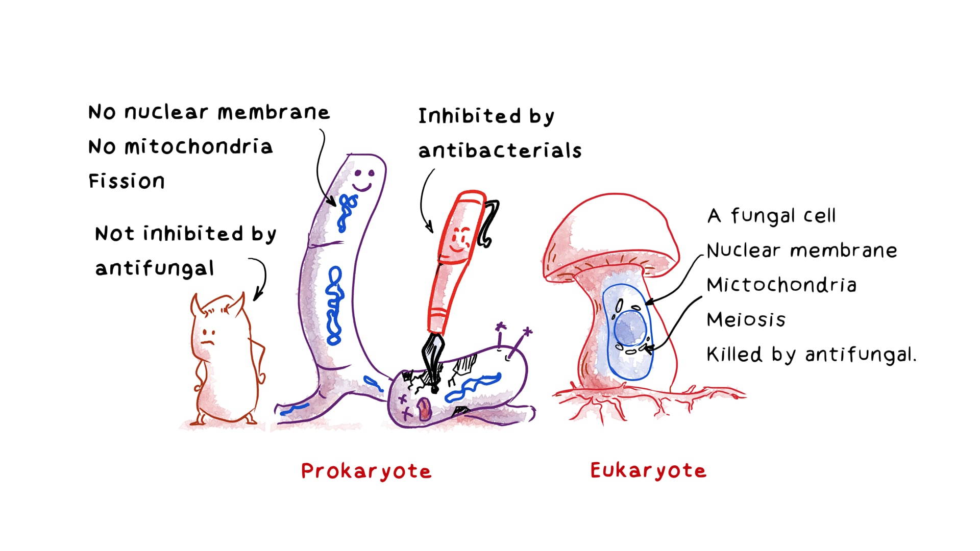
- * Covering the Gram +ive plasma membrane (phospholipid bi-layer), there are up to 60 peptidoglycan (PG) layers.
- * The gram -ive plasma layer is covered by up to three PG layers and an outer membrane containing lipopolysaccharides.
- * Beta-Lactamase is the most important factor in the periplasmic space, enzymes that can break down penicillins.
What happens to the gram stain?
- *The small molecules of dye get trapped in the PG layer of gram +ive bacteria and in both the outer membrane and the PG layers of the gram -ive bacteria.
- * At this stage both gram +ive and gram -ive look purple under the microscope.
- * When iodine (+) is added, it attaches to the molecules of dye (-) and creates a bigger molecule, which gets trapped in the PG layers. Color does not change.
- *When alcohol is added it dissolves the lipid portion and dehydrates the cells.
- * In the gram +ive cells the water is pulled out by the alcohol and the 60 layers are compressed, trapping the molecules of dye, keeping the purple color
- * In the gram -ive cells, the alcohol dissolves the more abundant lipids, and the three PG layers are too weak to trap the dye molecules so they escape, making the cells colorless.
NB: Using too much alcohol on the gram +ive cells will eventually break down the PG layers allowing the dye to escape so you have to be careful. - *The final step is to add safranin, a counterstain, that will turn the colorless gram -ive cells red, but will not be absorbed by the compressed gram +ive cells which stay purple.
And that is how gram staining works!
Learning objectives of this video are the following:
1. Review of the procedure of gram staining.
2. Results of gram staining in case of gram positive and gram negative bacteria.
3. How gram staining works?
Presented by Dr. Mobeen Syed
Following answers are created by ChatGPT. Occasionally the answer may be harmful, incorrect, false, misleading, incomplete, or limited in knowledge of world. Please contact your doctor for all healthcare decisions. Also, double check the answer provided by the AI below.
Faculty
In addition to the presenter, following authors may have helped with the content writing, review, or approval:
CME, CE, CEU and Other Credit Types:
ACCME Accreditation Statement
The DrBeen Corp is accredited by the Accreditation Council for Continuing Medical Education (ACCME) to
provide continuing medical education for physicians.
AMA Credit Designation Statement
The DrBeen Corp designates this enduring material for a maximum of 0.25 AMA PRA Category 1
Credits™.
Physicians should claim only the credit commensurate with the extent of their participation in the
activity.
Disclosure Information
In accordance with the disclosure policies of DrBeen Corp and the ACCME (Accreditation Council for
Continuing Medical Education), we are committed to upholding principles of balance, independence,
objectivity, and scientific rigor in all of our Continuing Medical Education (CME) and Continuing
Education (CE) activities. These policies include the careful management and mitigation of any relevant
financial relationships with organizations that are not eligible.
All members of the Activity Planning Committee and presenters have disclosed their relevant financial
relationships. The DrBeen Corp CE Committee has thoroughly reviewed these disclosures and determined
that these relationships are not deemed inappropriate in the context of their respective presentations.
Additionally, they are found to be consistent with the educational objectives and the integrity of the
activity.
| Faculty | Disclosures |
|---|---|
| Author declares no conflict of interest. |
Instructors
Microbiology
0.17 CME
Gram staining
10:05
Dr. Mobeen Syed
 0.50 CME
Bacterial Structure
23:09
Dr. Mobeen Syed
 0.20 CME
Bacteriophage
11:30
Dr. Mobeen Syed
 0.25 CME
Bacterial Genetic Recombination via Transduction
15:15
Dr. Mobeen Syed
 0.25 CME
Bacterial Genetic Recombination via Conjugation
16:30
Dr. Mobeen Syed
 0.17 CME
Bacterial Gene Recombination Via Transformation
09:47
Dr. Mobeen Syed
 0.23 CME
Bacterial Genetic Recombination via Transposons
13:33
Dr. Mobeen Syed
 1.00 CME
Antibiotics Overview
58:29
Dr. Mobeen Syed
 0.21 CME
Penicillin: Mechanism of Action
12:15
Dr. Mobeen Syed
 0.50 CME
Future Antivirals Using Bacterial Immune Sensors
26:02
Dr. Mobeen Syed
 0.75 CME
Dr. Steven Phillips Discusses Dementia and Chronic Diseases
42:55
Dr. Mobeen Syed
How Does Monkeypox Virus Work
27:08
Dr. Mobeen Syed
 0.25 CME
Why We Catch More Colds During The Cold Weather - Harvard Study
16:12
Dr. Mobeen Syed
Candida Auris is Spreading in the US - CDC Says
16:06
Dr. Mobeen Syed
 0.25 CME
Shingles Vaccine Shingrix - Indications, Mechanism, and Side Effects
22:26
Dr. Mobeen Syed
 0.25 CME
Elevated Interleukin 6 (IL-6) and Normal C Reactive Protein (CRP)
21:22
Dr. Mobeen Syed
 0.50 CME
Measles
26:15
Dr. Mobeen Syed
0.20 CME
Bacteriophage
11:30
Dr. Mobeen Syed
 0.14 CME
Alpha Hemolytic Streptococci
08:20
Dr. Mobeen Syed
 0.25 CME
Streptococcus Viridans
16:05
Dr. Mobeen Syed
 0.25 CME
Streptococcus Pneumoniae (Pneumococcus) - Pathophysiology and Management
21:53
Dr. Mobeen Syed
 0.25 CME
Pneumococcus (memory aid)
18:03
Dr. Mobeen Syed
 0.23 CME
Streptococcus Pyogenes - Clinical Foundations
13:42
Dr. Mobeen Syed
 0.25 CME
Streptococcus Pyogenes - Diseases and The Management Approach
20:12
Dr. Mobeen Syed
 0.23 CME
Streptococcus Agalactiae
13:23
Dr. Mobeen Syed
 0.17 CME
Streptococcus Bovis
09:46
Dr. Mobeen Syed
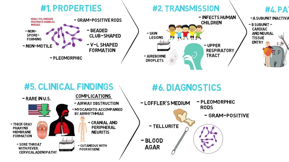
Corynebacterium Diptheriae
03:39
Dr. Mobeen Syed
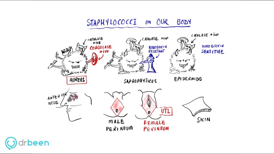
Staphylococci Properties, Diseases, Diagnosis, and Management Approach
24:45
Dr. Mobeen Syed
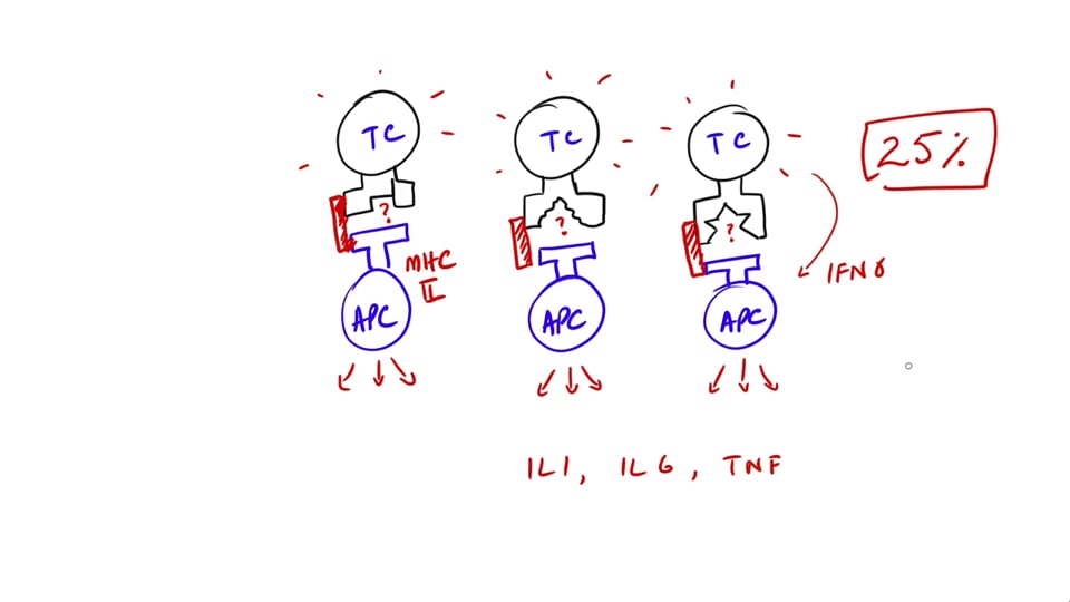
Why do superantigens cause cytokine storm?
09:08
Dr. Mobeen Syed
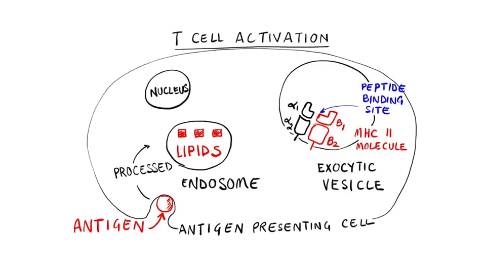
Staphylococcus - Superantigens
13:50
Dr. Mobeen Syed
 0.25 CME
Staphylococcus - Toxic Shock Syndrome and Its Management
15:37
Dr. Mobeen Syed
Staphylococcal Scalded Skin Syndrome
13:00
Dr. Mobeen Syed
Staphylococcal Gastroenteritis
06:42
Dr. Mobeen Syed
Staphylococcal Infective Endocarditis - (Staphylococcus)
11:49
Dr. Mobeen Syed
Staphylococcus - Diagnosing Acute Infective Endocarditis (Modified Duke's Criteria)
11:26
Dr. Mobeen Syed
 0.13 CME
Current Approaches to Acute Infective Endocarditis (Staphylococcus Series)
07:23
Dr. Mobeen Syed
 0.25 CME
Staphylococcus Skin Infections Impetigo Folliculitis Furuncles or Boils Carbuncles Cellulitis Surgical Wound
16:55
Dr. Mobeen Syed
Dr. Mobeen Syed
 0.50 CME
Bacillus Anthracis - Properties and Pathophysiology
33:31
Dr. Mobeen Syed
 0.75 CME
Anthrax - Types, Signs and Symptoms, Labs, Prophylaxis, Treatment, Vaccination, and Prognosis
40:07
Dr. Mobeen Syed
0.50 CME
Future Antivirals Using Bacterial Immune Sensors
26:02
Dr. Mobeen Syed
 0.17 CME
Bacillus Cereus
09:43
Dr. Mobeen Syed
 0.50 CME
Clostridium Tetani
23:56
Dr. Mobeen Syed
 0.50 CME
Clostridium Botulinum
28:07
Dr. Mobeen Syed
 0.25 CME
Clostridium Perfringens
21:00
Dr. Mobeen Syed
 0.25 CME
Clostridium Difficile
21:59
Dr. Mobeen Syed
 0.25 CME
Listeria Monocytogenes
21:09
Dr. Mobeen Syed
 0.21 CME
Actinomyces and Actinomycosis
12:26
Dr. Mobeen Syed
 0.24 CME
Nocardia and Nocardiosis
13:56
Dr. Mobeen Syed
 0.50 CME
Taenia Solium (Pork Tapeworm)
33:11
Dr. Mobeen Syed
 0.50 CME
Taenia Saginata (Beef Tapeworm)
23:52
Dr. Mobeen Syed
0.50 CME
Measles
26:15
Dr. Mobeen Syed
Write A New Comment
2 Comments
naveedkashif7@*.com
Jan 13 2023, 8:16 pm
Very nice and informative
attaulhadiii@*.com
Feb 14 2020, 1:22 am
Which method is used for staining mycobacteria?