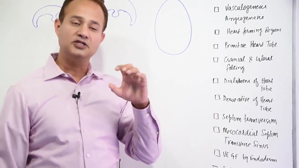
Difference between intra-embryonic mesoderm and coelom Vasculogenesis angiogenesis Heart forming regions and the development of the heart tube Heart tube dilatations and their derivatives Septum transversum mesocordial septum transverse sinus Retinoic acid Levo Dyenin VE GF Kartagener's syndrome/primary cilliary disease Situs inversus.
STUDY NOTES:
HEART TUBES
INTRODUCTION: Cardiovascular system is the first organ system to start developing and reach a functional state; which is even before its own development is complete. Cardiovascular development occurs during the 3rd to 5th week of intraembryonic life. Up until the second week, diffusion is enough for embryo to receive the oxygen, nutrition and to get rid of the waste products. The lacunae of maternal blood filled spaces and embryonic villi of the syncytio-trophoblast are involved in an intimate relationship, which allows gaseous exchange via diffusion in between the developing embryo and the maternal blood. However, into the third week, diffusion alone is not sufficient to match the needs of the growing embryo and it needs a circulatory system, hence heart has to develop.
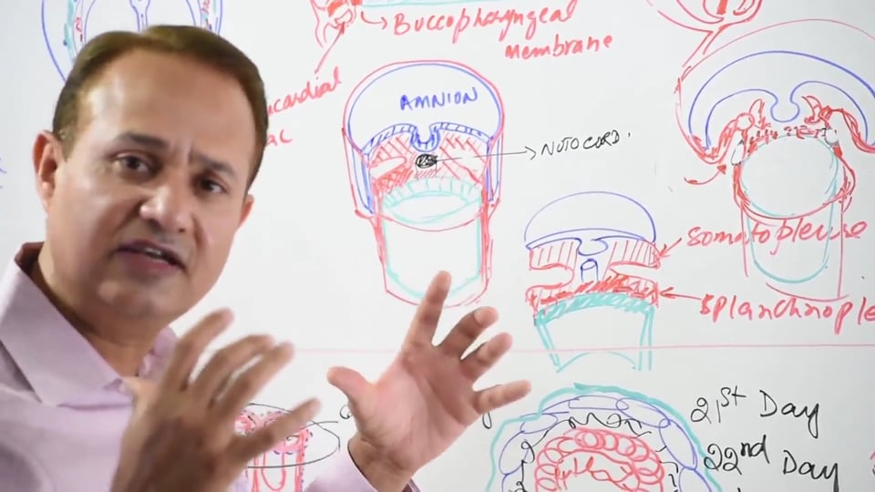
GASTRULA STAGE: Gastrula is a stage of development of embryo, when it is in the form of tri-laminar germ disc including ectoderm, mesoderm and endoderm. Endoderm is associated with umbilical vesicle; ectoderm is associated with Amniotic cavity. Cytotrophoblast around the gastrula develops multiple cavities. The cavities merge to form one big cavity called extraembryonic coelom. The cytotrophoblast is connected to embryo through the connecting stalk. The cellular layer from cytotrophoblast which covers the gastrula is called extraembryonic mesoderm. The Gastrula is a slipper shaped structure. From the dorsal view it has a neural plate with a primitive node a primitive pit and a primitive groove. Cranial side has a Buccopharyngeal membrane which would later develop into mouth. Caudal end has a cloacal membrane which would develop into anus.
HEART FORMING REGIONS: Around 15th day of intraembryonic life, multiple cavitations start appearing in the lateral plate mesoderm which later merge together to form a Horseshoe-shaped Intraembryonic Coelom. The Horseshoe shaped Coelom divides lateral plate mesoderm into two layers which are called Splanchnopleure (connected to underlying Endoderm) and Somatopleure (connected to overlying Ectoderm). It’s the splanchnic layer of mesoderm which mainly forms the cardiogenic area in the latter half of the 3rd week. In addition to this, Neural crest cells also contribute to heart formation especially the Aorticopulmonary septum and the Endocardial Cushion regions.
Around 17the day of intraembryonic life, the Endoderm layer of the Gastrula secretes VEGF, which causes Ectodermal cells to migrate into the cranial end of the underlying mesoderm and form Blood islands. A horseshoe shaped area forms on either side of the neural plate. The blood islands formed above the Prechordial plate (Cranial side) are called PrimaryCardiogenic or Heart-forming regions.
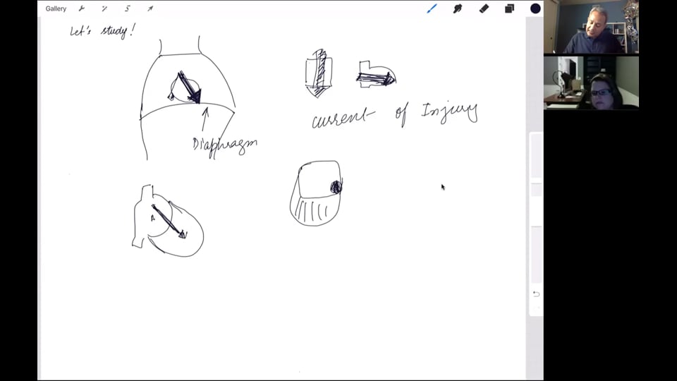
CRANIAL & LATERAL BODY FOLDINGS: By the 17-18 days brain starts developing at a faster pace and overtakes the body’s growth so that the heart descends downwards to the chest region. Cephalic-caudal Body folding ensures that the cardiogenic area and septum Transversum (future diaphragm) come to lie under and below the prechordal plate and in frontof the foregut. The heart hangs from the foregut by a connection called prechordium.Lateral body folding approximates the intraembryonic coelom in the midline, folds of which give rise to adult pericardial sacs which envelope the cardiogenic area in the midline.
DEVELOPMENT OF PRIMITIVE HEART TUBES: The ectodermal cells migrate into the mesoderm as cardiogeniccells which condense to form a pair of primordial Heart tubes. The pharyngeal area mesoderm contributes further cells which form a secondary heart forming region around the primordial heart tubes. Further cells are contributed the Splanchnic Mesoderm which form the myocardium around the primordial heart tubes. This newly formed Myocardium will start secreting Hyaluronic acid and other connective tissue components which are termed together and called as Cardiac Jelly. Cardiac jelly in future becomes the connective tissue of the heart. These newly formed primordial heart tubes are surrounded by pericardial cavity which provides the outer Parietal layer of the Pericardium which is adherent to the Fibrous pericardium in the adult heart. The Caudal or Inflow part of the Heart tube that is Sinus Venosus provides the cells which form the visceral or inner layer of pericardium, also called EPICARDIUM. By the 21st day the two primordial heart tubes fuse by apoptosis into a single endocardial or Heart tube. On 22nd day the embryonic heart starts beating.
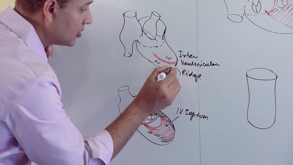
DILATATIONS & DERIVATIVES OF THE HEART TUBE: The Primordial heart tube now orients itself into a cephalic INFLOW (Venous region) and a cranial OUTFLOW (Arterial Region) ends. At this point the primitive heart tube has five dilatations which are as following:
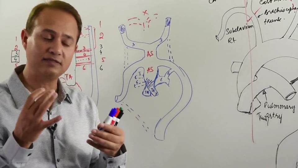
- Truncus Arteriosus (Arterial Outflow region): Forms Adult Aorta, Pulmonary trunk and their respective semilunar valves.
- Bulbus Cordis: Forms Smooth parts of Adult right ventricle (conus arteriosus) and left ventricle (aortic vestibule).
- Primitive Ventricle: Forms trabeculated/rough parts of right and left ventricles.
- Primitive Atrium: Forms trabeculated/rough parts of right and left atriums i.e., the pectinate muscles.
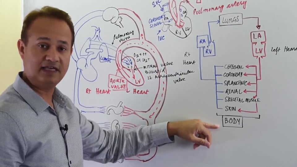
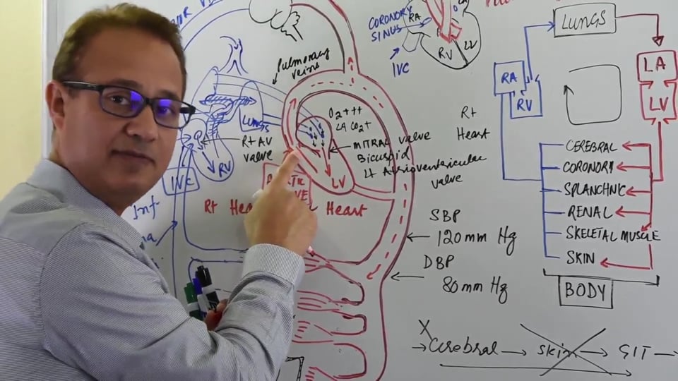
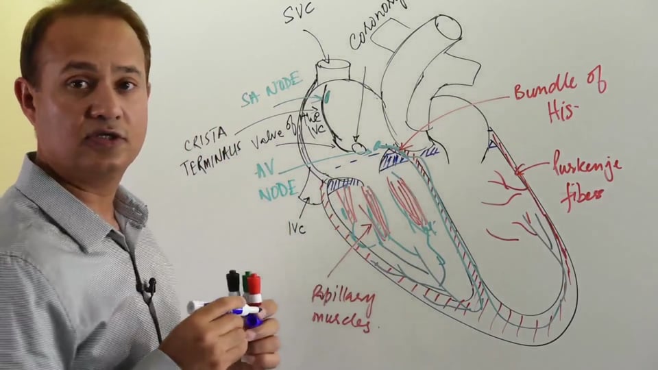
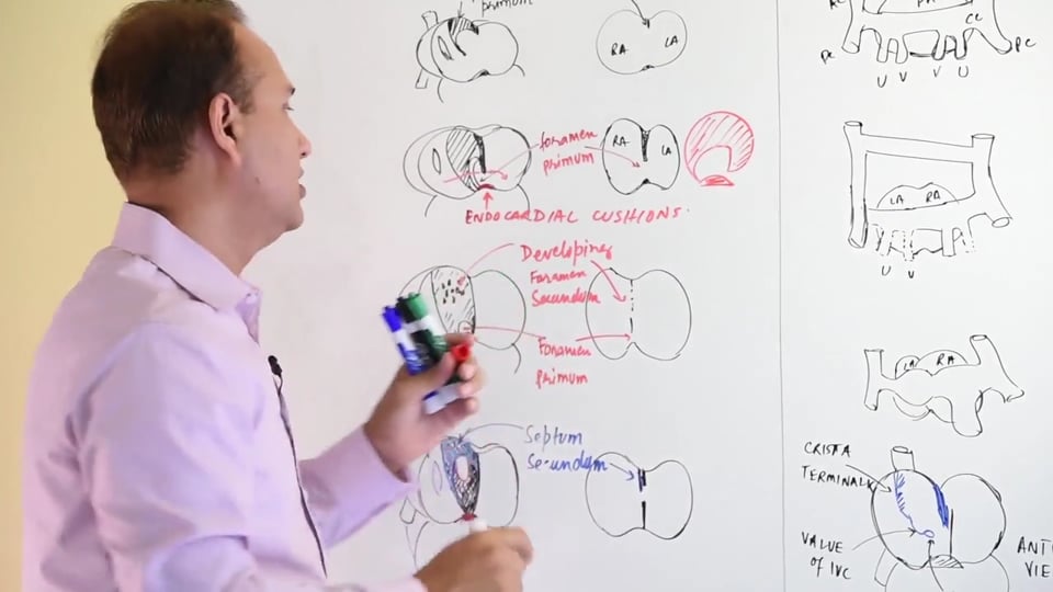
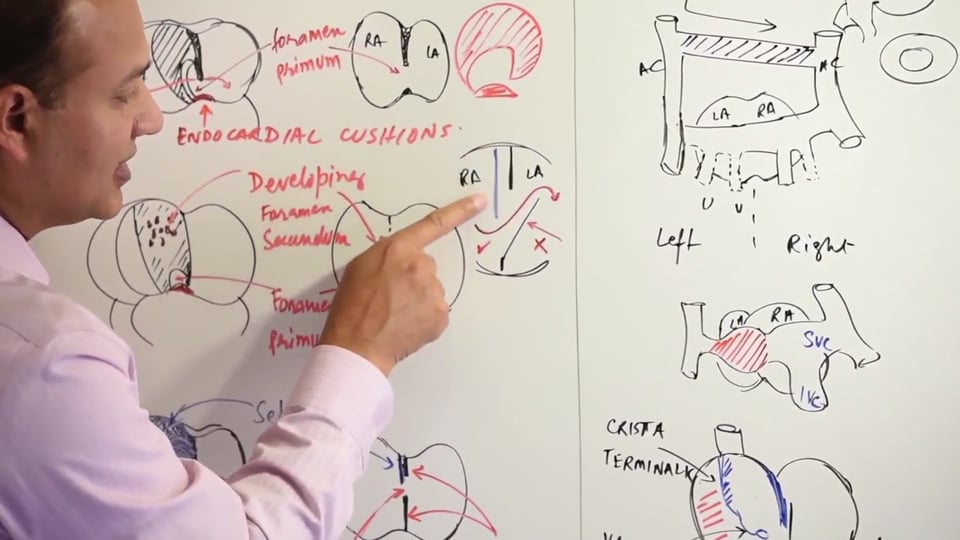
- Sinus Venosus: On the right side it forms Sinus Venarum (smooth part of right Atrium), Superior vena cava and the inferior vena cava. On the left side it forms Coronary sinus and oblique vein of left atrium.
Note: (a) The vascular parts when incorporated into the adult heart form the smooth regions of the heart whereas the primitive chambers form the rough or trabeculated parts of the respective adult heart chambers.
(b) Incorporation of parts of the Pulmonary veins forms the smooth-walled part of the left Atrium. On the right side, incorporation of right sinus venosus forms the smooth-walled part of right atrium.
DEXTRAL LOOPING: The heart tube at this point undergoes Right sided bending or rotation which referred to as Dextral looping. The Truncus Arteriosus or the ventricularend of the heart tube grows more rapidly and tends to fold downwards, forwards and to the right side. Subsequently, the lower parts of the tube i.e., the primitive atria and sinus venosus tend to fold upwards, backwards and to the left side. This dextral looping tends to place the chambers of heart in their adult anatomic positions where the right ventricle forms most of the right border plus the anterior surface of the heart and the left atrium is the posterior most chamber of the heart. Also, the ventricles are rather more anteriorly placed relative to atria in an adult heart.
THE ROLE OF LEVO-DYNEIN, DEXTROCARDIA, SITUS INVERSUS & KARTEGENER SYNDROME: Levo-Dynein is a protein which involved in the formation of Cilia. However, Levo-Dynein also functions to create symmetry within the human body. An abnormality of Levo-dynein can lead to symmetry problems such as, Situs Inversus whereas the visceras tend to be present on the opposite sides of their normal anatomical location. It can also lead to Dextrocardia, which is a rare clinical condition in which the Apex of the heart is located on the right side of the body. The above two abnormalities often present as part of Kartagener Syndrome (Primary Ciliary Dyskinesia). Kartagener Syndrome results due to a defect in the dynein arm of the cilia which renders cilia immotile. It is a cause of infertility in both males and females due to immotile sperm and dysfunctional fallopian tube cilia respectively. In females there's an additional risk of ectopic pregnancies. Besides Dextrocardia on CXR and infertility in both sexes, Kartagener Syndrome can also lead to Bronchiectasis and recurrent sinusitis due to ineffectiveness of mucociliary escalator.
MESOCORDIUM & TRANSVERSE SINUS: Post the cranial-caudal body folding, the embryonic heart tubes come to lie in front of the foregut. This is before the fusion of the primordial heart tubes into a single Heart tube. At this point the primordial heart tubes are connected to the foregut via the Mesocordium. The Mesocordium itself is a derivative of peritoneum. Subsequently, a gap appears within the Mesocordium which is called transverse sinus and this eventually results in degeneration of Mesocordium, following which the Pericardial cavity is thus separated from the Foregut.
TERATOGENS EXPOSURE: Teratogens are substances (normally drugs) which can either cause birth defects or they can accelerate other embryonic deformities that are present. Developing embryo is most susceptible to teratogens exposure during its embryonic period which is from 3rd to 8th week (first 2 months).This is because the embryonic period is the time when most organ systems are developing; hence teratogen exposure at this point can be disastrous. Teratogens frequently tested by the USMLE are given below:
- ACE Inhibitors: Cause Renal damage
- Aminoglycosides: Cranial nerve 8, Vestibulocochlear Nerve Abnormalities.
- Carbamazepine: Facial dysmorphism, developmental delay and neural tube defects.
- Lithium (used to treat the manic phase of Bipolar disorder): Ebstein Anomaly in which the tricuspid valve leaflets are displaced inferiorly into the right ventricle. It presents with widely split S2 and Tricuspid Regurgitation.
- Phenytoin: Can cause fetal Hydantoin syndrome i.e., cleft palate, cardiac defects and phalanx or fingernail hypoplasia.
- Tetracyclines: Discoloration of teeth.
- Thalidomide: Limb defects.
ROLE OF RETIONOIC ACID: Increased retinoic acid concentration in an area of blood vessel formation leads to formation of a venous channel. However, a decrease in retinoic acid concentration favours the formation of an arterial channel.
DIFFERENCE BETWEEN VASCULOGENESIS & ANGIOGENESIS: Vasculogenesis is defined as formation of a new blood vessel literally from nothing or scratch. Angiogenesis on the other hand is defined as, formation of a new blood vessel from an existing vascular channel by branching. Regions of an existing blood vessel bud of as part of angiogenesis to create a new branch.
In this lecture Dr. Mobeen will discuss:
1. Introduction to cardiovascular system embryology. (3:30)
2. Vasculogenesis/ Angiogenesis in the heart. (5:57)
3. Formation of the cavity of the heart. (7:00)
4. Mesodermal components of the heart. (15:36)
5. Cranial and lateral folding. (17:39)
6. Formation of the pericardial sac. (21:44)
7. Formation of the heart tube. (36:00)
8. Formation of the layers of the heart. (41:36)
9. Truncus arteriosus and ventricle cavities. (44:29)
10. Folding of the tube. (47:00)
11. Dextrocardia and kartagener syndrome. (51:50)


Write A New Comment
2 Comments
zeeshanpmdc@*.com
Jun 11 2025, 10:49 am
can able download it
drrasheedk@*.com
Dec 05 2023, 4:43 pm
I could not recieve the zoom link "drrasheedk@yahoo.com."